For most of human history, the first few weeks of pregnancy have been a complete mystery. We know that a sperm fertilizes an egg, and nine months later, a baby is born. But the crucial moment when the embryo attaches to the mother's womb—known as implantation—happens deep inside the body, hidden from view. Doctors often call this period the "black box" of human development. 1,2
It is a dangerous time. Statistics show that 30% to 60% of all conceptions fail before a woman even knows she is pregnant, and about 10% of known pregnancies end in miscarriage 1. Most of these failures happen during implantation. Because we cannot ethically or safely watch this happen inside a human body, scientists have been working in the dark.
Until now.
A wave of groundbreaking research is changing everything. Scientists are now building synthetic embryos (made from stem cells) and artificial endometrial models (lab-grown uterine linings) to recreate this hidden moment in a petri dish. These tools are not just cool science experiments; they are windows into the very beginning of life, offering new hope for treating infertility and preventing miscarriages.
This article explores how these models are built, what they have revealed about the physical and chemical "handshake" between mother and embryo, and the ethical questions arising from creating life-like structures in the lab.
Part 1: The "Seed" — Creating Synthetic Embryos
To study implantation, you need an embryo. For decades, researchers relied on surplus embryos donated by patients undergoing In Vitro Fertilization (IVF). While incredibly valuable, these embryos are scarce, often of lower quality, and subject to strict ethical rules that usually prevent studying them beyond 14 days. 3
Enter the "Blastoid."
Blastoids are stem cell-based embryo models (SEMs). Instead of needing a sperm and an egg, scientists use pluripotent stem cells—master cells that can turn into any tissue in the body. By giving these stem cells specific chemical signals, they self-organize into structures that look and act remarkably like a 5-day-old human embryo, or blastocyst. 4
What Makes a Blastoid?
A natural blastocyst has three main parts, and a good blastoid must mimic them all:
- The Epiblast: These cells will become the fetus itself.
- The Trophoblast: These cells form the outer shell and will eventually become the placenta. Their job is to attach to the uterus.
- The Hypoblast: These cells will form the yolk sac, which nourishes the early embryo.
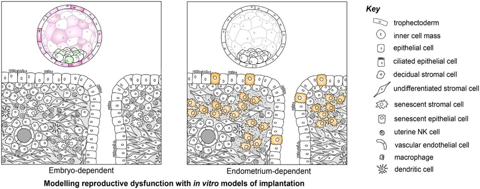
--Image of: --Comparison of Blastoid and Natural Embryo Figure 1: The evolution of embryo models. Scientists have moved from simple clusters of cells to complex "blastoids" that mimic the structure of a natural embryo. These can be combined with endometrial models to study implantation. Source: Rawlings et al., 2026.
However, these models are not perfect. They don't develop into full human beings, and they often lack the precise organization of a natural embryo. But for studying the mechanics of attachment, they are a game-changer. They allow scientists to perform experiments that would be impossible with real embryos, such as testing how specific genes affect the ability to attach to the womb.
Part 2: The "Soil" — Engineering the Artificial Womb
An embryo needs a place to land. The lining of the uterus, called the endometrium, is complex. It has a surface layer of epithelial cells (the "skin" of the uterus) and a deep layer of stromal cells (the structural tissue). It also contains glands that secrete "uterine milk" to feed the embryo, and immune cells that decide whether to accept or reject the pregnancy. 10,11
Recreating this in a lab has been a massive bioengineering challenge.
The "Inside-Out" Problem
Early attempts to grow endometrial tissue created organoids—tiny, 3D balls of tissue. While useful, they had a major flaw: they grew "apical-in." The sticky surface that an embryo is supposed to attach to (the apical side) was trapped on the inside of the sphere. It was like trying to land a plane on a runway that has been rolled up inside a tube.
To fix this, scientists developed "apical-out" organoids. By changing the chemical environment, they flipped the cells so the sticky, receptive surface faced outward.
The Mouse Model Success
In a recent study, Japanese researchers created a breakthrough mouse endometrial organoid. They mixed epithelial cells (surface) and stromal cells (deep tissue) and let them self-organize. Remarkably, the cells sorted themselves out: epithelial cells formed a shell on the outside, and stromal cells filled the inside, perfectly mimicking the structure of the uterine lining. 13,15
Crucially, this model didn't use an artificial gel (like Matrigel) to hold everything together. This allowed the embryo to touch the cells directly, just as it would in the body 12. When they added hormones to mimic pregnancy, the organoid formed "pinopodes"—tiny protrusions that help trap the embryo. 16
The Human Solution: CREST
For human research, scientists at Stanford and Cambridge developed a system called CREST (Cell-engineered Receptive Endometria ScaffoldTechnology). 1
Instead of relying on self-organization alone, they built a scaffold. They encapsulated human endometrial stromal cells inside a specialized hydrogel (a water-rich jelly) reinforced with collagen proteins to make it stiff, like real tissue 17. Then, they plated epithelial cells on top.
The result was a piece of lab-grown tissue that acted exactly like a receptive uterus. It responded to hormones like estrogen and progesterone. The stromal cells underwent decidualization—a process where they change shape and start secreting proteins to support a pregnancy 18,19. The epithelial surface even developed glands that secreted nutrients, proving the tissue was functional.
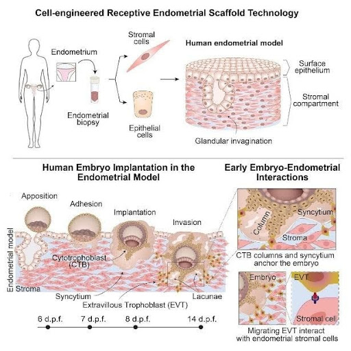
--Image of: --The CREST Model Figure 1,2: The CREST model recreates the human endometrium. It features a surface layer for the embryo to attach to and a deep stromal layer for it to invade. Source: Molè et al., 2026. 20
Part 3: The First Handshake — When Embryo Meets Womb
With both the "seed" (embryo/blastoid) and "soil" (endometrial model) ready, scientists could finally observe the moment of contact.
The Invasion
In the CREST model, scientists watched as human embryos attached to the surface. Within 24 hours, the embryos hatched from their protective shells. By day 8, they had flattened and begun to burrow 21,22. By day 10, they were gone—completely buried inside the lab-grown tissue, just as they would be in a mother's womb. 22
This experiment revealed specific "handshakes" between the cells. For example, the trophoblast cells (from the embryo) and the stromal cells (from the uterus) communicate using a signaling pathway called PROS1-AXL. PROS1 is a protein signal sent by the embryo, and AXL is the receiver on the uterine cells. When scientists blocked this signal with a drug, the embryos stopped growing and failed to invade properly 23,24. This suggests that some cases of infertility might be caused by a failure in this specific molecular conversation.
The Mouse vs. Human Difference
Using these models, researchers have also highlighted how different we are from mice. In the mouse organoid model, the embryo attaches and the uterine cells actively wrap around it, a process called entosis. The mouse embryo doesn't just push in; the uterus pulls it in.
In humans, the process is more invasive. The human embryo acts like a parasite, aggressively digging into the tissue. The CREST study showed human embryos sending out "columns" of cells to anchor themselves deep into the matrix, establishing the roots of the placenta. 26
Part 4: The Physics of Life — Embryos Use "The Force"
We often think of biology as chemistry—hormones, proteins, and genes. But a fascinating study published in Science Advances 27 shows that implantation is also a mechanical process. Embryos physically pull themselves into the womb.
Using high-resolution imaging and special sensors, researchers measured the traction forces exerted by embryos. They found that human embryos are surprisingly strong.
Tug-of-War
When a human embryo touches the uterine lining, it doesn't just stick; it pulls. The study found that human embryos generate "foci" of traction—specific spots where they grip the tissue and pull radially, like tightening a drawstring bag 28. This pulling force remodels the collagen matrix around them, stiffening the tissue to create a stable anchor. 29
Mouse embryos behave differently. They exert anisotropic forces, meaning they pull in specific directions rather than uniformly. This directional pulling might help orient the mouse embryo correctly in the uterus, ensuring its head and tail develop in the right direction. 30
Mechanosensitivity: The Embryo Can "Feel"
Perhaps the most incredible finding is that embryos are mechanosensitive. They can "feel" how stiff or soft the uterus is, and they respond to external forces. 28,31
In one experiment, researchers poked the matrix near a mouse embryo with a microneedle to simulate external pressure (like a uterine contraction). The embryo responded by changing its growth direction, pointing its body axis toward the source of the force 31. Human embryos responded by sending out cell protrusions toward the pressure, actively trying to grab onto the source of the stimulation. 32
This means the uterus isn't just a passive bed; its stiffness and movements (contractions) physically guide the embryo, telling it where to attach and which way to grow. If the uterus is too stiff (perhaps due to scarring) or too soft, the embryo might not be able to "grip" properly, leading to implantation failure. 33,34
Part 5: Ethical Frontiers — Creating Life-Like Entities
As these models become more realistic, they enter a gray area of ethics. If a synthetic embryo in a lab-grown womb has a beating heart (which happens around day 22 in nature), is it a person?
Currently, international guidelines, such as those from the European Society of Human Reproduction and Embryology (ESHRE), distinguish between "natural" embryos and "embryo-like structures" (ELS). 35
The Status of the "Synthetic"
The consensus is that current synthetic embryos are not equivalent to natural ones. They lack the full potential to grow into a baby because they don't develop the placenta and yolk sac perfectly 36. Therefore, they are not granted the same moral status as a natural embryo.
However, ESHRE warns that this could change. If a synthetic embryo passes the "Turing test" of biology—meaning it is indistinguishable from a real embryo and could theoretically result in a live birth if transferred to a womb—it should be treated with the same strict ethical rules. 37
The 14-Day Rule
For decades, scientists have agreed not to grow human embryos in the lab beyond 14 days (the point where the individual identity is set and the nervous system begins to form) 38. But because synthetic embryos are not "real" embryos, they technically fall outside this law in many countries. 39
Bioethicists are now debating whether to extend this limit to 28 days. 40 Extending the limit would allow scientists to study the formation of organs and the early heart, which are prone to congenital defects. However, it raises profound questions about the moral value we assign to developing human life. 41
Currently, researchers using the CREST model or blastoids stop their experiments before the 14-day mark or when specific developmental milestones are reached, to stay well within ethical boundaries. 42
Conclusion: A New Era of Reproductive Science
We are witnessing the end of the "black box" era of human reproduction. By combining synthetic embryos with engineered endometrial tissues, scientists have created a window into the most secretive week of human life.
We now know that implantation is a dynamic dance. It involves:
- Chemical conversations: Signals like PROS1-AXL ensuring the embryo and mother are compatible. 23
- Physical struggles: Embryos actively pulling on the womb to anchor themselves. 43
- Structural reorganization: The uterus building a "nest" of glands and blood vessels to feed the new life. 11
The potential benefits are immense. These models could allow doctors to test drugs for safety during early pregnancy without risking a real fetus 44. They could help diagnose why some women suffer recurrent miscarriages by testing their own endometrial cells against standard embryo models 45. They might even lead to non-hormonal contraceptives that work by simply preventing that first, crucial handshake. 20
As we peel back the layers of mystery surrounding our own origins, we must tread carefully, balancing scientific curiosity with ethical responsibility.
But for the millions of people struggling to conceive, these tiny, lab-grown clusters of cells represent a massive beacon of hope.